Thermal Images: Teeth

The Thermogram Center visualizes acute dental findings in ~ 1 in 3 clients.

Thermal Images: Teeth

Click here for Silent Killers in the Mouth

Click here for root canals

Click here for cavities that can lead to abscesses

Click here for teeth extractions that can lead to cavitations

Click here for the tooth organ chart

Click here for Tirza Derflinger’s “Correlations between Thermal Imaging, 2D X-ray and 3D Cone Beam Tomography with Symptomatic Relief After Surgery.”

Thermal Findings and Failed Root CanalsThermal Findings and Failed Root Canals

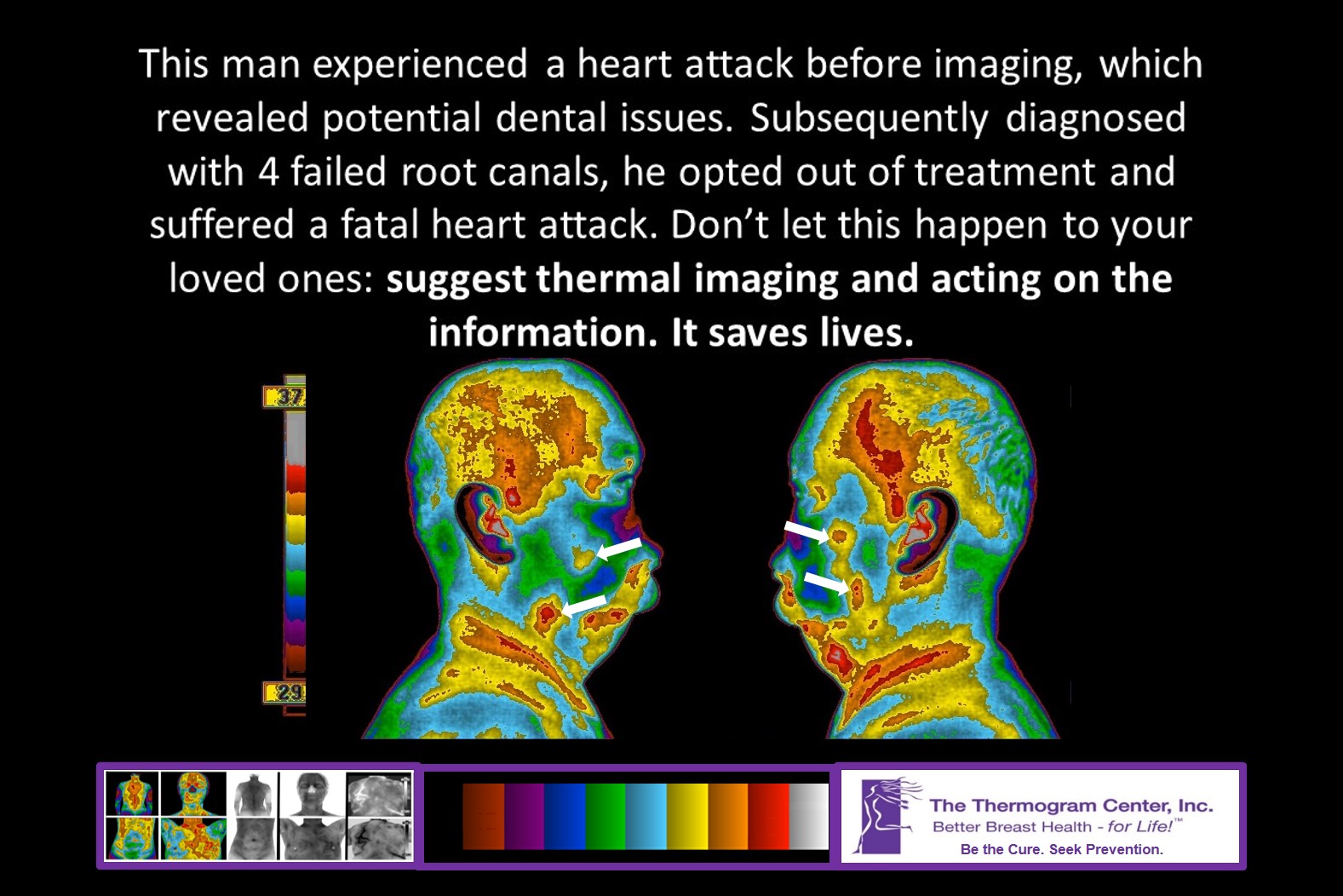
This 72 year old male had experienced innumerable root canals and decades of dental work which was completed in 1998. In 2007 he suffered a heart attack.

When thermally imaged in 2009, he was free of symptoms or concerns for his teeth and therefore surprised when his thermal imaging raised the index of suspicion for potentially acute dental issues associated with previously root canaled teeth, subsequently diagnosed by a dental professional.

Since certain dental and bone infections can lead to heart attacks, auto-immune disease and other life threatening conditions, these findings were significant and potentially crucial to his longevity.

Before and After Effects of Failed Root Canal Extraction on the Breasts

Before and After Effects of Failed Root Canal Extraction on the Breasts

Top Row – 2012: (Former Camera)

Note the lymph flow from the neck into the chest and breasts, particularly the right breast. R – TH 4, L – TH 2

Bottom Row – 2015: (Current Camera)

Note the significant reduction in chest inflammation, the increased symmetry between breasts, and the lack of heat/thermal features therein. R – TH 1, L – TH 1

Following the left tooth #14 extraction, the client’s Thermobiological Risk in the right breast decreased from HIGH risk (4) to LOWEST risk (1). Prevention was supported!!! Of what, we’ll never know…

Acute Thermal Findings Lead to 4 Cavitation Diagnoses

Acute Thermal Findings Lead to 4 Cavitation Diagnoses

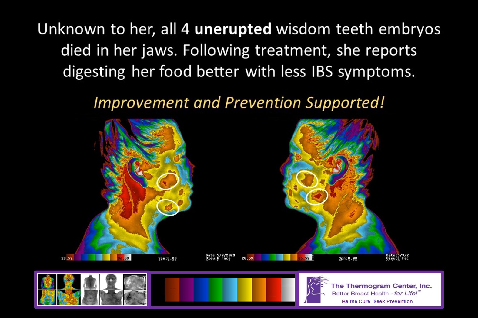
03-18-21 imaging of this 43 year old client revealed acute thermal findings in all four back quadrants of the jaws. The most acute is seen in the lower left jaw.

Subsequent 3D CBT diagnosed cavitations where all four wisdom teeth were removed at ~20 years of age.

Cavitation treatment starts 05-03-21, and with it, prevention of potential health issues that may have otherwise resulted, ie autoimmune conditions, heart attacks and cancer.

Worsening Thermal Findings Leads to Diagnosis of Abscess in Tooth #31

Worsening Thermal Findings Leads to Diagnosis of Abscess in Tooth #31

TOP ROW: 2017Notice the normal, cold nose; cool sinus regions; and warm spot under the gum line of the lower right mid-to-back teeth. Since under-the-gum line issues/infections typically grow very slowly due to drastically reduced blood supply under the gum line, the client was recommended to repeat face imaging in 3-5 years.

BOTTOM ROW: 2020Notice that the nose and sinus regions are warming, and that the spot under the gum line of the lower right mid-to-back teeth is worsening. Since under-the-gum line finding worsened, diagnostic evaluation was warranted.

Subsequent 3D Cone Beam Tomography  diagnosed an abscess of tooth #31 in spite of the client not having any jaw symptoms. Since issues with this tooth may contribute to heart attack and breast disease, prevention is supported.

Thermal Signs Consistent with Potential Dental Issues

Thermal Signs Consistent with Potential Dental Issues

This 64 year old female had her upper wisdom teeth removed in the mid 1980s and the lowers removed in 2008.

When thermally imaged in 2010, she was free of symptoms or concerns for her teeth. However, the thermal imaging raised the index of suspicion for dental issues and cavitation.

Further evaluation ensued with a Doctor of Dental Science and this client was diagnosed with cavitation with each wisdom tooth removed, requiring cavitation surgery and bone grafting. (The lower left wisdom tooth extraction site had the least cavitation).

Thermal Findings Warranting Further Evaluation

This female had no concerns regarding her oral health. Note the significant heat/thermal finding at “B”, which is consistent with an active lymph node, among other things. In the event of an underlying tooth issue, she sought dentistry to determine the cause of this finding. Dental xray of tooth #18 indicated abnormality at “C”, consistent with infection. This tooth had previously undergone root canal and was crowned. Months of treatment to improve the health of the tooth proved unsuccessful, and the tooth was extracted. After extraction, a contributing factor/likely cause of infection was observed: a long/vertical fracture/crack at “D”. Her dentist found soft tissue infection between the roots on the opposite side of the tooth at “E”.

With some infections, thermal findings may be present, raising the index of suspicion. Because each tooth is connected to other body parts/organs, and infections can effect these body parts/organs, it is important to not have dental-related infections. For instance, some can cause heart attacks; all 8 back teeth are connected by meridian to the heart. As far as this client knows, she removed the infection at tooth #18 before it impacted corresponding body parts, including her left lung and large intestine.

Dental Issues and Breast Cancer

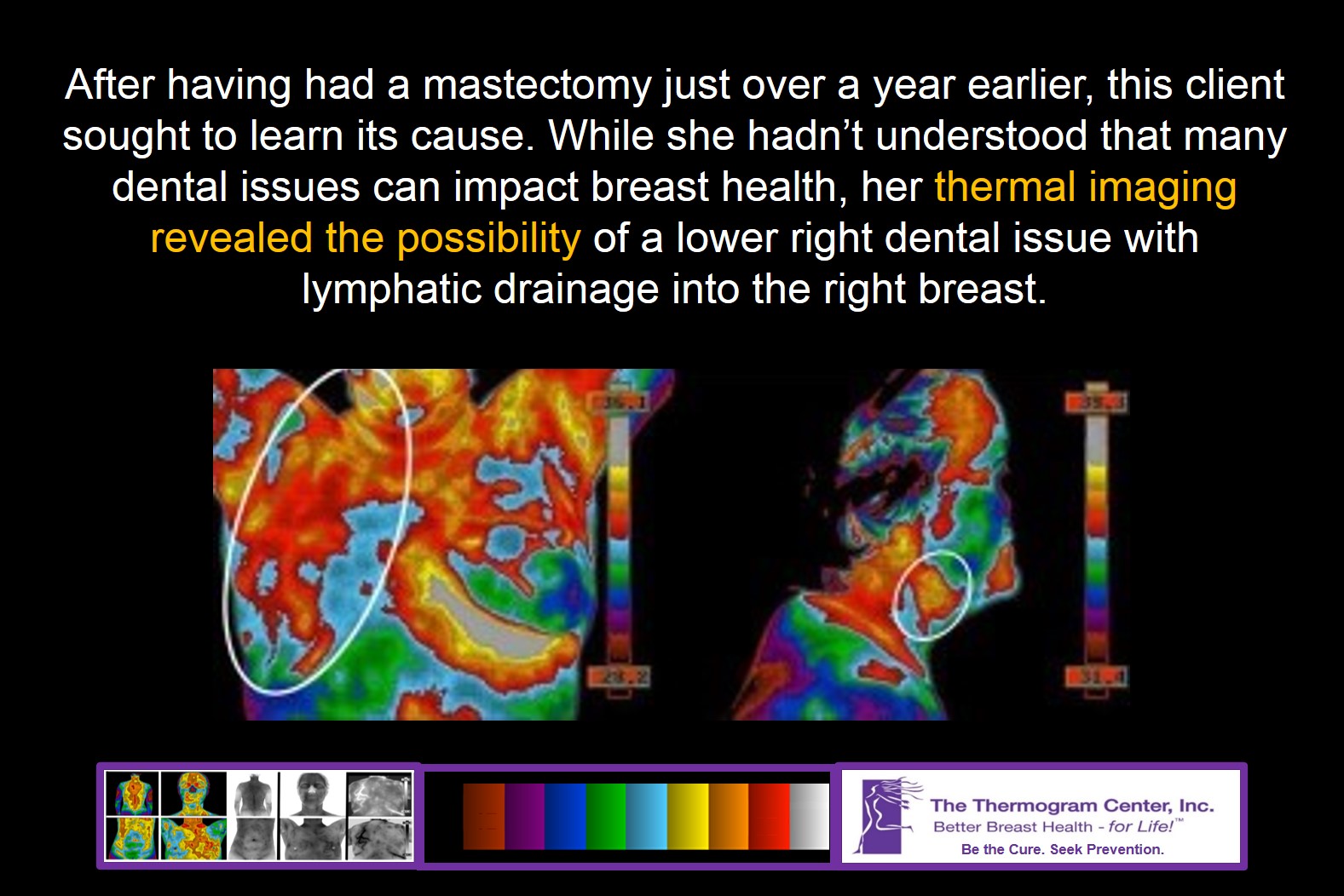
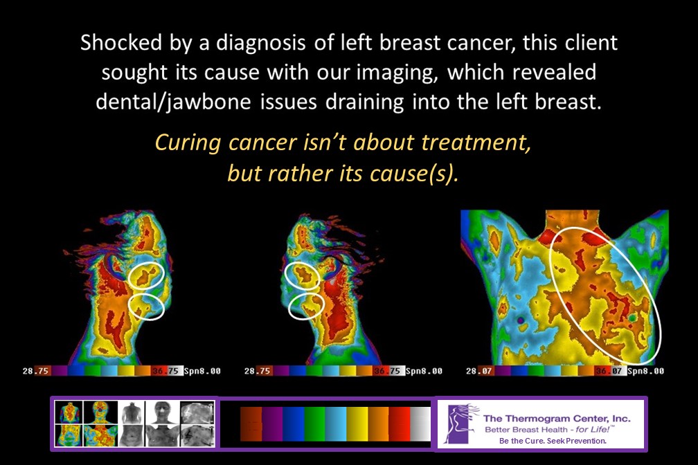
Dental Issues and Breast CancerThis 46 year old female used thermal imaging in 2010 after having had a mastectomy just over a year earlier. While she hadn’t understood that many dental issues can affect the breast, her thermal imaging and the tooth and organ chart revealed to her the possibility of a lower back molar issue and breast connection. In the upper left image, lymphatic drainage from the right side of the mouth into the chest is apparent.


Thermal Findings and Cavitations

Thermal Findings and CavitationsThis 54 year old female reported depleting energy levels and concern for developing auto-immune disorders if the source of her chronic fatigue could not be resolved.

Her thermal imaging raised the index of suspicion for potential dental issues and infection with lymphatic involvement/drainage into the chest. Note the excess heat and thermal patterns along the left chest. While her follow-up tests were normal for the breast, her dentist diagnosed 2 cavitations in her right jaws – where wisdom teeth had been extracted twenty years earlier.

Since cavitations can compromise the immune system and overall health, she is now working to resolve this known condition.


Tooth Connected to Lung Issue?


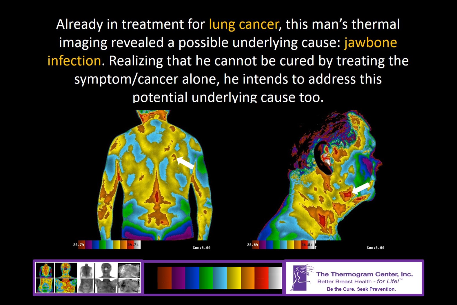
Tooth Connected to Lung IssueThis male had experienced pulmonary embolisms weeks prior to full-body imaging, which consisted of 24+ images. While most of the images were absent of findings, the thermal findings in his anterior neck view raised the index of suspicion for oral infection with lymphatic drainage from the left lower jaw. The client reported that he had a tooth worked on there in recent months, but that it still “wasn’t right”. (The tooth is energetically linked (by meridian) to the lungs.)


Thermal Analyses + Symptoms Lead to Scrutiny, Diagnosis, and Treatment


Thermal Analyses + Symptoms Lead to Scrutiny, Diagnosis, and TreatmentThis 45 year old female had a complaint with an upper right front tooth. Her dental hygienist indicated that all was well.

During an imaging appointment in 2008, thermal analyses raised the index of suspicion for mild oral infection.

Still symptomatic and armed with her thermal imaging report, she requested that her dentist conduct a thorough exam.

The dentist found that the tooth was cracked and that infection had developed under the tooth that required care and a crown.

She later reported that the tooth feels much better.